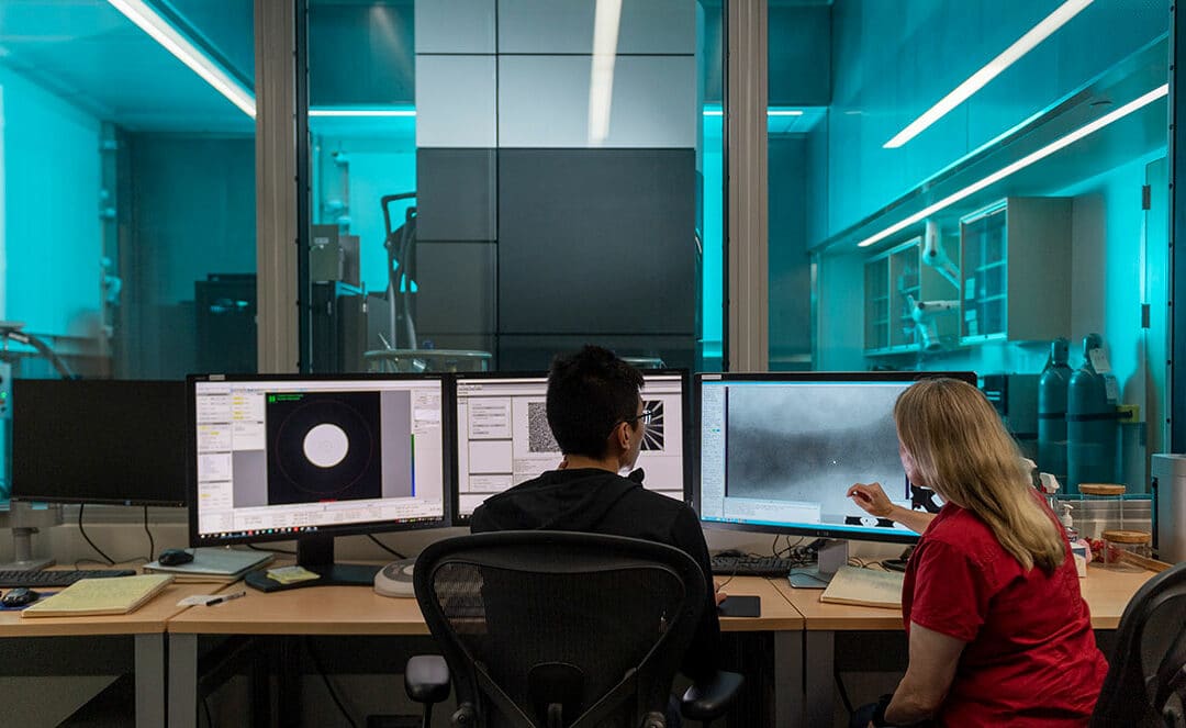
Microscopes have come a long way from what many people remember from their high school biology class. Instead of peering through a lens, scientists can now create 3-D images by shooting beams of electrons at protein structures that have been frozen to hold their shape.
This state-of-the-art technology, called cryo-electron microscopy or cryo-EM, drives a promising collaboration between the UW-Madison Department of Biochemistry and the Morgridge Institute. Recognizing they risked falling behind, the partners worked to build a center that supports a broad range of research on campus and is now a national hub for training and research development.
Cryo-EM is all about getting clearer, more detailed images of the structure of molecules. Scientists need this atomic-level resolution to understand, for example, how proteins malfunction with disease and how to target them when developing a new drug. The specimen is flash-frozen in liquid ethane, which helps reduce the damage that inevitably occurs every time the electrons hit to secure an image.
“We’re pioneering different aspects of thinking about computation, hardcore algorithm development, and how we use these microscopes to look at challenging biological questions.”Elizabeth Wright
The technology dates from 1974, but the field began to boom about ten years ago when more advanced hardware hit the market. Like many universities in the country, UW-Madison realized it needed to get serious about cryo-EM or risk falling seriously behind. Investigators at Morgridge and the biochemistry department started inviting the world’s best experts to campus to give talks so they could learn how it works and understand what they needed to do.
In 2018, the field took off in the United States when the National Institutes of Health developed three national cryo-EM centers for single-particle analysis, a sort of plug-and-play approach where a scientist wants a single image to look at a specimen.
UW-Madison decided to establish its own center, focused on the more complex approach called tomography. Similar to a person undergoing an MRI, the specimens are rotated and 120-140 images are taken from every angle to reconstruct the molecule.
Morgridge and the biochemistry department also joined forces in 2018 to recruit Elizabeth Wright from Emory University, where she had built a similar Cryo-EM center from the ground up. Everything fell into place when the NIH put out a proposal to support cryo-EM tomography centers – UW-Madison applied and won.
The depth of the partnership has been clear from the start. Buying expensive equipment through a state university can require jumping through hoops that can take as long as two years, so the department offered to take on other expenses if Morgridge could secure the microscopes. “We did all the negotiating afterwards, because we trusted each other enough to know that it was going to work, and it did,” says Brad Schwartz, CEO of Morgridge.
After heavy renovations, including pouring thick concrete slabs topped by an antivibration table to keep the sensitive equipment still, the first four microscopes were speedily delivered just as the pandemic forced the campus to close in March in 2020.
Ten years ago, scientists were looking at what Wright calls “blobology,” when all the pictures taken looked like little blobs of molecules and you couldn’t exactly see what’s going on. The images of today, by contrast, let you see where all the atoms are positioned – like moving from play dough to tinker toys.
This process generates so much data that normal laptops can’t handle the computation required to sort and analyze all the images. This is where the high-throughput computing capacity of Morgridge comes into play, what has become as essential to a lab as lighting. “Without really good computation, the cryo-EM microscopes are just over overpriced paperweights,” says Wright.
Brian Bockelman, a research computing investigator at the institute, draws on deep experience working with physicists on projects requiring massive amounts of data because the fundamentals are the same. His computational infrastructure is helping the cryo-EM center extract the particles that interest a researcher to generate the higher-resolution 3-D structures they need. “I would love it if researchers on campus drowned us with computational and data challenges so they can really start pushing us to think better,” Bockelman says.
Morgridge also brings the expertise of Tim Grant, a biomedical imaging specialist and assistant professor of biochemistry, who develops algorithms and applies new methods for improving the imaging and its analysis.
The center has talked to a vast number of investigators, and is already working with nearly 50 groups spanning many different types of science. In addition to the researchers studying proteins, many are looking at viruses such as SARS-CoV-2 and HIV. Others work with bacteria to understand how to develop new antimicrobials and antibiotics. Some groups want to look more at tissue, such as the eye, or brain tissue to understand different neurodegenerative diseases and brain development.
“We’re pioneering different aspects of thinking about computation, hardcore algorithm development, and how we use these microscopes to look at challenging biological questions,” says Wright.